|
From rxpgnews.com Breast
Apropos of National Breast Cancer Awareness month, researchers with the U.S. Department of Energy�s Lawrence Berkeley National Laboratory (Berkeley Lab) have created a first-of-its-kind model for studying how breast tissue is shaped and structured during development. The model may shed new light on how the misbehavior of only a few cells can facilitate metastatic invasion because it shows that the development of breast tissue, normal or abnormal, is controlled not only by genetics but also by geometry. Though created specifically for the study of breast tissue, this model should also be applicable to the study of tissue development in other organs as well.
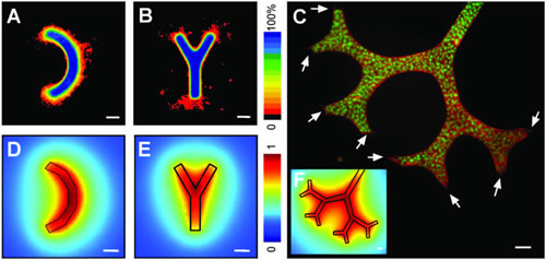
The paper is entitled: Tissue Geometry Determines Sites of Mammary Branching Morphogenesis in Organotypic Cultures. Co-authoring the paper with Bissell were Celeste Nelson and Jamie Inman, both members of Bissell�s research group, and Daniel Fletcher and Martijn VanDuijn, from the University of California at Berkeley�s Bioengineering Department. Fletcher also holds an appointment with Berkeley Lab�s Physical Biosciences Division. In mammals, breast tissue begins to morph into milk glands at the onset of puberty. In this process, called branching morphogenesis, epithelial cell tubes begin to migrate outward, invading the surrounding pad of fat cells to form a widely branched tree of milk ducts. Branching morphogenesis is known to involve a complex interplay of both intracellular and extracellular signals that within the context of the tissue determines precisely where new branches are initiated. Said co-author Nelson, a post-doctoral bioengineer who will soon have her own research group at Princeton University, �One group of cells within a tubule is instructed to form a branch or to bifurcate, whereas a neighboring group is not. While branching morphogenesis is common to many organs, including the lung, kidney and salivary gland, we still do not have a precise understanding of how spatial positioning is determined. Given that the mammary ductal network branches out from pre-existing epithelial tubules, we hypothesized that the position of cells within a tubule might provide contextual information to instruct branch site initiation.� Bissell is one of the leading proponents of the idea that a cell�s genetic information is supplemented by contextual information encoded within the microenvironment that surrounds the cell. To define the role of positional context, she and Nelson developed a 3-D micropatterned assay for mammary epithelial branching morphogenesis. This assay enabled them to control the initial geometry of epithelial tubules and to quantify the positions at which they branched. In their studies, Bissell, Nelson and their colleagues engineered epithelial tubules of defined geometry by embedding functionally normal mouse mammary epithelial cells in cavities of a collagen gel. The epithelial cells formed hollow tubules, according to the size and shape of the collagen cavities. These tubules began branching out into the gel within 24 hours after being treated with epidermal growth factor. To quantify branching and to represent its magnitude and position, the researchers stained the cell nuclei with fluorescent dye and imaged them using confocal microscopy. �We confirmed that the position of branching depended on the initial geometry of the tubule,� said Nelson. �Increasing the length of the tubules increased the magnitude of branching, although cells still branched exclusively from the ends. Curved tubules branched preferentially from the convex side of the curve. Asymmetric branching was also observed in bifurcated tubules and trees, which preferentially branched from distal positions.� The process of normal branching morphogenesis is precise and quantitative, but invasionary; when something goes wrong the process may lend itself to metastasis. With this demonstration of how the normal function of branching morphogenesis is controlled, Bissell believes researchers can now look for ways in which faulty tubule geometry leads to malignancy. �In breast cancer, it is most often metastasis rather than the primary tumor that kills a patient,� said Bissell. �We have learned something really dramatic about the regulation of normal branching morphogenesis and this should help us understand how and why things go wrong. Our next step is to put pre-malignant cells � cells that are already losing their way but are not yet malignant � into our model and see what happens. When we do, perhaps this will provide us with new ideas for intervening and preventing pre-malignant cells from becoming fully malignant.� All rights reserved by www.rxpgnews.com |