Nerve tissue interfaced with a computer chip
Jun 3, 2006, 01:15, Reviewed by: Dr. Priya Saxena
|
|
The scientists in Martinsried developed a revolutionary noninvasive technique that enables them to record neural communication between thousands of nerve cells in the tissue of a brain slice with high spatial resolution.
|
By Max Planck Institute of Biochemistry,
For the first time, scientists at the Max-Planck Institute for Biochemistry in Martinsried near Munich coupled living brain tissue to a chip equivalent to the chips that run computers.
Before informational input perceived by the mammalian brain is stored in the long-term memory, it is temporarily memorised in the hippocampus. Understanding the function of the hippocampus as an important player in the memory process is a major topic of current brain research. Thin slices of this brain region provide the appropriate material to study the intact neural network of the hippocampus.
Methods commonly used in neurophysiology are invasive, restricted to a small number of cells or suffer from low spatial resolution. The scientists in Martinsried developed a revolutionary noninvasive technique that enables them to record neural communication between thousands of nerve cells in the tissue of a brain slice with high spatial resolution. This technique involves culturing razor-thin slices of the hippocampus region on semiconductor chips. These chips were developed in collaboration with Infineon Technologies AG and excel in their density of sensory transistors: 16384 transistors on an area of one square millimeter record the neural activity in the brain.
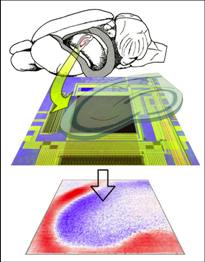 |
| Figure: A thin tissue slice of a rat hippocampus region (top) is cultivated on a semiconductor chip with 16.384 sensory transistors per square millimetre (center, dark coloured square). Following excitation the chip maps the electrical activity of the neurons (bottom), caused by activity of synapses (red: positive, blue: negative). Copyright: Max Planck Institute of Biochemistry |
The researchers under Peter Fromherz have reported this news in the online edition of the Journal of Neurophysiology (May 10, 2006).
Recording the activity patterns of the united cell structure of an intact mammalian brain tissue represents a significant technological breakthrough. Employing the new technique, the biophysicists working under the direction of Peter Fromherz were able to visualize the influence of pharmaceutical compounds on the neural network. This makes the �brain-chip� from Martinsried a novel test system for brain and drug research.
As early as 1991, Peter Fromherz and his co-workers succeeded in interfacing single leech nerve cells with semiconductor chips. Subsequent research gave rise to bidirectional communication between chip and small networks of a few molluscan nerve cells. In this project, it was possible to detect the signalling between cells via their synapses. The chips used in these studies were developed and produced by the scientists themselves. The production requirements of the chip described above made collaboration with industry indispensable. With the resulting novel hybrid system of neural tissue and semiconductor, the scientists take a great step forward towards neurochip prosthetics and neurocomputation. 
- M. Hutzler, A. Lambacher, B. Eversmann, M. Jenkner, R. Thewes, and P. Fromherz: Highresolution multi-transistor array recording of electrical field potentials in cultured brain slices. Journal of Neuropyhsiology. Preprint online (May 10, 2006). doi:10.1152/jn.00347.2006
www.biochem.mpg.de
The Max Planck Institute of Biochemistry in Martinsried near Munich was founded in 1973 as the successor of the three formerly independent institutes MPI of Biochemistry (founded in 1912 as Kaiser-Wilhelm-Institut in Berlin-Dahlem), MPI for Protein and Leather Research (founded in 1954 in Regensburg) and the MPI for Cell Chemistry (founded in 1956 in Munich). Research at the institute focuses on studies of the relationship between the structure and the activity of biological systems with various degrees of complexity, i.e. nucleic acids as carriers of the genetic information and proteins or peptides as structural components of the three-dimensional organization of the cell. Their folding and function is likewise the subject of the research such as cell organells, membranes, viruses, oncogenes, archaebacteria, higher cells and whole organisms. These serve as models for the investigation of the molecular bases of biological and pathophysiological phenomena: Cell-cell interactions, cell division, cell growth and - differentiation, gene regulation, signal transmission by nerves, photosynthesis, evolution and cancer. A special strength of the institute is the various methodical expertise in the areas of protein chemistry, molecular biology and structure analysis. Research in scientific departments and working groups is supported by central service facilities for DNA- and protein sequence analysis, DNA- and peptide-synthesis, NMR and mass spectrometry, a central library, an information retrieval and managing center, a computing center with bioinformatics unit, an animal house and central workshops. New methods in molecular biology and genetic engineering led to the development of a biomedically oriented basic research of molecular medicine. In this field of research medical questions are connected with molecular-biological techniques, which opens promising possibilities in the pathogenesis research and the development of new diagnostic and therapeutical approaches. The institute cooperates in this area with the pharmaceutical industry on national and international level. Several research groups of the institute are working on this medical orientated field of research, others are dealing with pure basic research.
|
For any corrections of factual information, to contact the editors or to send
any medical news or health news press releases, use
feedback form
Top of Page
|