
|
|
First-ever Images of Living Human Retinas Sprung a Surprise
Oct 27, 2005, 00:32, Reviewed by: Dr.
|
|
"Over time, we were able to shift their natural perception of yellow in one direction, and then the other. This is direct evidence for an internal, automatic calibrator of color perception. These experiments show that color is defined by our experience in the world, and since we all share the same world, we arrive at the same definition of colors."
|
By University of Rochester,
First-ever images of living human retinas have yielded a surprise about how we perceive our world. Researchers at the University of Rochester have found that the number of color-sensitive cones in the human retina differs dramatically among people�by up to 40 times�yet people appear to perceive colors the same way. The findings, on the cover of this week's journal Neuroscience, strongly suggest that our perception of color is controlled much more by our brains than by our eyes.
"We were able to precisely image and count the color-receptive cones in a living human eye for the first time, and we were astonished at the results," says David Williams, Allyn Professor of Medical Optics and director of the Center for Visual Science. "We've shown that color perception goes far beyond the hardware of the eye, and that leads to a lot of interesting questions about how and why we perceive color."
Williams and his research team, led by postdoctoral student Heidi Hofer, now an assistant professor at the University of Houston, used a laser-based system developed by Williams that maps out the topography of the inner eye in exquisite detail. The technology, known as adaptive optics, was originally used by astronomers in telescopes to compensate for the blurring of starlight caused by the atmosphere.
Williams turned the technique from the heavens back toward the eye to compensate for common aberrations. The technique allows researchers to study the living retina in ways that were never before possible. The pigment that allows each cone in the human eye to react to different colors is very fragile and normal microscope light bleaches it away. This means that looking at the retina from a cadaver yields almost no information on the arrangement of their cones, and there is certainly no ability to test for color perception. Likewise, the amino acids that make up two of the three different-colored cones are so similar that there are no stains that can bind to some and not others, a process often used by researchers to differentiate cell types under a microscope.
Imaging the living retina allowed Williams to shine light directly into the eye to see what wavelengths each cone reflects and absorbs, and thus to which color each is responsive. In addition, the technique allows scientists to image more than a thousand cones at once, giving an unprecedented look at the composition and distribution of color cones in the eyes of living humans with varied retinal structure.
Each subject was asked to tune the color of a disk of light to produce a pure yellow light that was neither reddish yellow nor greenish yellow. Everyone selected nearly the same wavelength of yellow, showing an obvious consensus over what color they perceived yellow to be. Once Williams looked into their eyes, however, he was surprised to see that the number of long- and middle-wavelength cones�the cones that detect red, green, and yellow�were sometimes profusely scattered throughout the retina, and sometimes barely evident. The discrepancy was more than a 40:1 ratio, yet all the volunteers were apparently seeing the same color yellow.
"Those early experiments showed that everyone we tested has the same color experience despite this really profound difference in the front-end of their visual system," says Hofer. "That points to some kind of normalization or auto-calibration mechanism�some kind of circuit in the brain that balances the colors for you no matter what the hardware is."
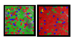 |
| Images of living human retinas showing the wide diversity of number of cones sensitive to different colors. (Photo credit: University of Rochester) |
In a related experiment, Williams and a postdoctoral fellow Yasuki Yamauchi, working with other collaborators from the Medical College of Wisconsin, gave several people colored contacts to wear for four hours a day. While wearing the contacts, people tended to eventually feel as if they were not wearing the contacts, just as people who wear colored sunglasses tend to see colors "correctly" after a few minutes with the sunglasses. The volunteers' normal color vision, however, began to shift after several weeks of contact use. Even when not wearing the contacts, they all began to select a pure yellow that was a different wavelength than they had before wearing the contacts.
"Over time, we were able to shift their natural perception of yellow in one direction, and then the other," says Williams. "This is direct evidence for an internal, automatic calibrator of color perception. These experiments show that color is defined by our experience in the world, and since we all share the same world, we arrive at the same definition of colors." 
- The findings are on the cover of this week's journal Neuroscience
www.rochester.edu
Williams' team is now looking to identify the genetic basis for this large variation between retinas. Early tests on the original volunteers showed no simple connection among certain genes and the number and diversity of color cones, but Williams is continuing to search for the responsible combination of genes.
|
For any corrections of factual information, to contact the editors or to send
any medical news or health news press releases, use
feedback form
Top of Page
|
|
|
|